How to
Use
How to
Use- Health & Beauty Protective Items Health & Medical Beauty & Personal Care
- Bags, Shoes & Accessories Luggage, Bags & Cases Shoes & Accessories
- Apparel, Textiles & Accessories Apparel Textile & Leather Product Fashion Accessories Timepieces, Jewelry, Eyewear
- Electronics Industrial Computer & Accessories Home Appliance Consumer Electronic Security & Protection
- Electronic Equipment, Component & Telecoms Electronic Equipment & Supplies Telecommunication
- Home, Lights & Construction Construction & Real Estate Home & Garden Lights & Lighting Furniture
- Gifts, Sports & Toys Gifts & Crafts Toys & Hobbies Sports & Entertainment
- Agriculture & Food Agriculture Food & Beverage
- Auto & Transportation Automobiles & Motorcycles Transportation
- Machinery, Industrial Parts & Tools Machinery Industrial Parts & Fabrication Services Tools Hardware Measurement & Analysis Instruments
- Metallurgy, Chemicals, Rubber & Plastics Minerals & Metallurgy Chemicals Rubber & Plastics Energy Environment
- Packaging, Advertising & Office Packaging & Printing Office & School Supplies Service Equipment
- K-Service Innotech Contents ICT Technology/Engineering
- ETC ETC


ITEM SPECIFICS
-
Brand
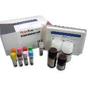
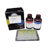
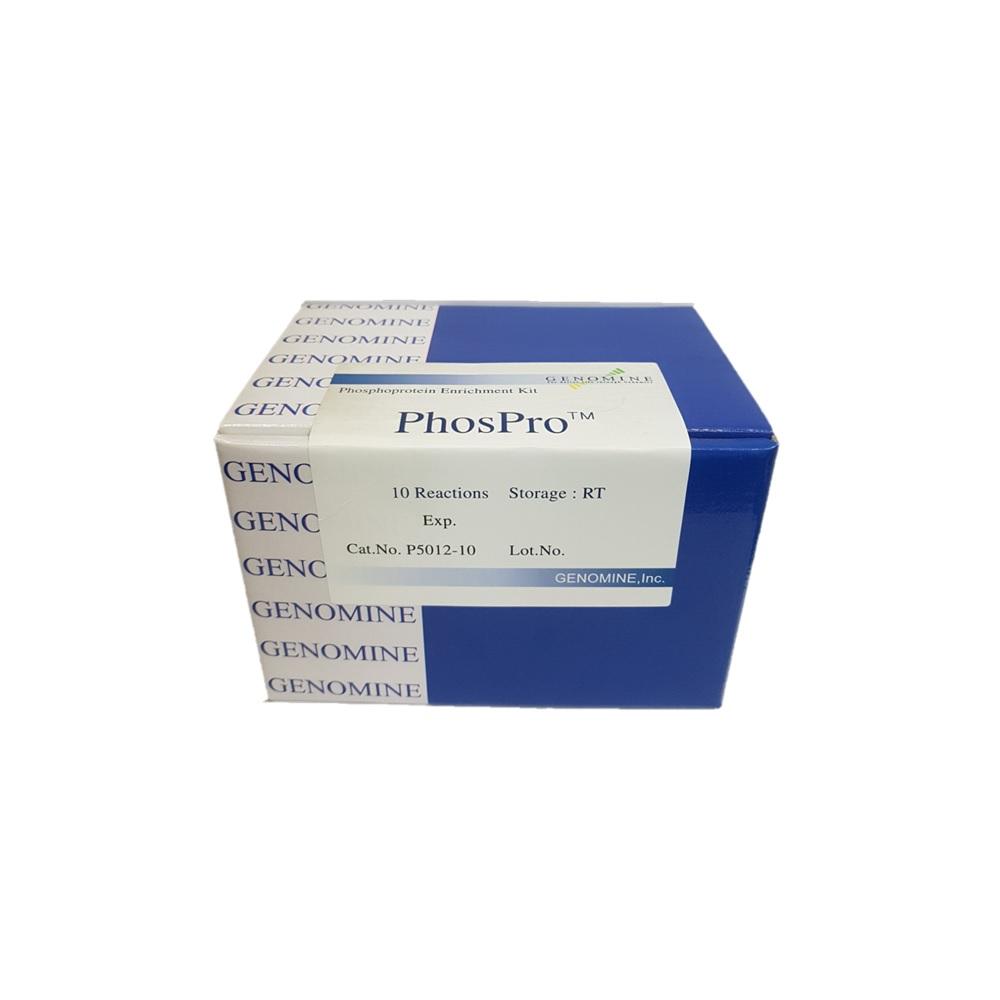
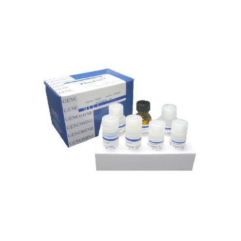
Antibody biotin conjugation kit
-
origin
Republic of Korea
-
Size(Capacity)
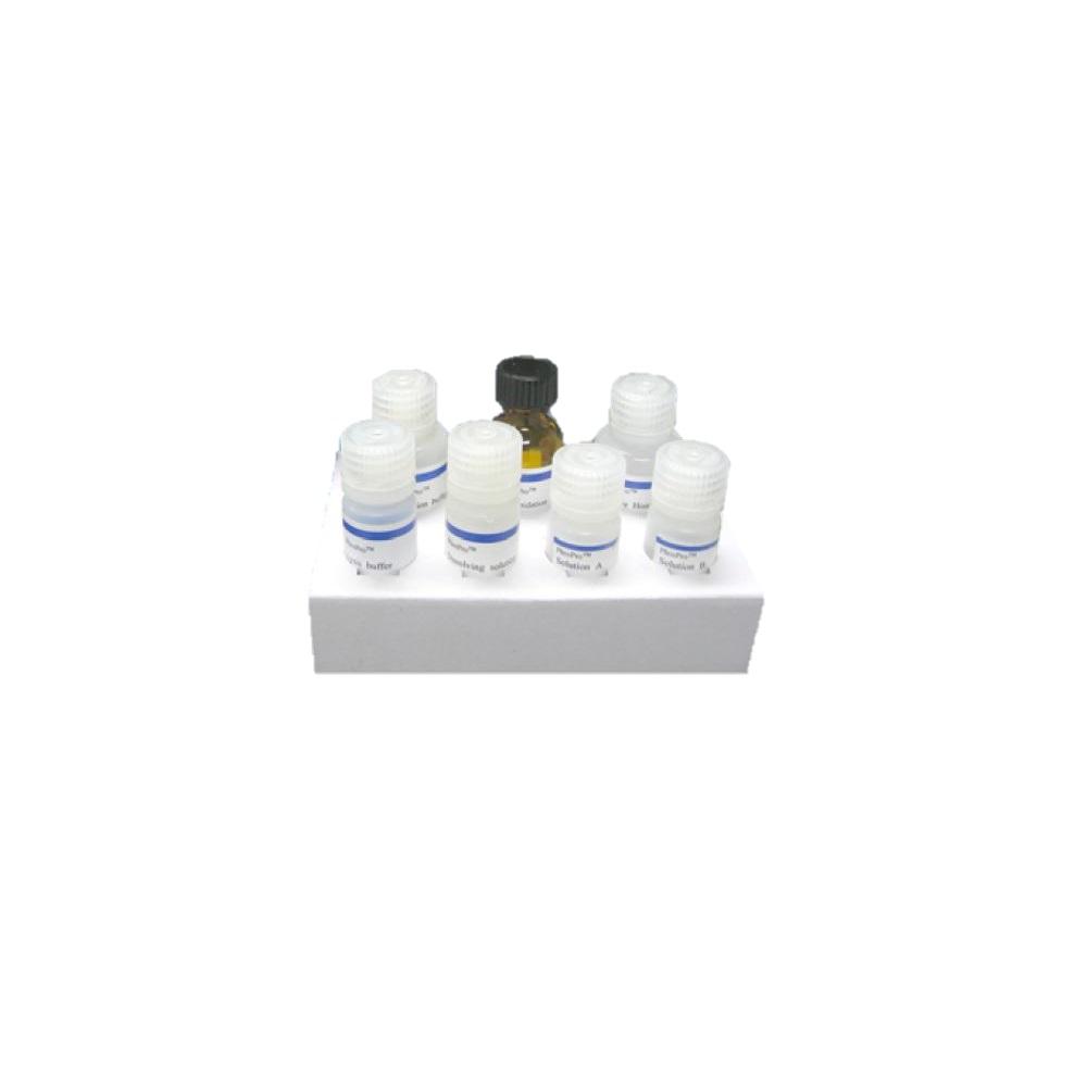
10 Reactions
PRODUCT DESCRIPTION
Protein phosphorylation is one of the most frequently occurred posttranslational modifications and plays a critical role in cellular regulatory events. Most cellular processes are in fact regulated by the reversible phosphorylation of proteins on serine, threonine and tyrosine residues. In fact, phosphorylation of proteins plays a key role in oncogenesis, cell signaling, apoptosis and immune disorders1. Despite the importance and widespread occurrence of this modification, profiling of phosphoproteins in cells is still a challenge, due to the low copy of phosphorylated proteins in cell and the relative amount of phosphoproteins compared to unphosphorylated proteins.
Radiolabeling by 32P labeling is frequently used conventional method for investigation of phosphoprotein profile in conjunction with 2-DE or 1-D gel electrophoresis and autoradiogram. Alternatively, western blot analysis probed by phosphoprotein-specific antibody is also used for this purpose.
Mass spectrometry has been shown to be a reliable and routine tool to identify proteins in a high throughput manner. However, the identification of phosphorylation by mass spectrometry is not a trivial matter and to this day is not routine also due to the low copy of phosphorylated proteins in cells.
This phosphoprotein enrichment and exclusion of unphosphorylated proteins provides advanced chance in detecting protein phosphorylation in gels with non-radiolabeling method(eg. Staining with fluorescence dye) and enables quantitative comparison between cells.
This phosphoprotein enrichment and exclusion of unphosphorylated proteins provides advanced chance in detecting protein phosphorylation in gels with non-radiolabeling method(eg. Staining with fluorescence dye) and enables quantitative comparison between cells.
PAYMENTS DETAILS
This supplier supports payments for offline orders
- WK
- Name : Sera Park
SHIPPING
Shipping from :
Republic of Korea
- 327 Saecheonnyeon-daero Nam-gu, Pohang-si, Gyeongsangbuk-do (37663)
GENOMINE,INC
The person in charge
Dong Su KimAddress
327 Saecheonnyeon-daero Nam-gu, Pohang-si, Gyeongsangbuk-do (37663)
Introduction
GENOMINE corporation is a biotech company dedicated to develop technologies and products of in-vitro diagnostics field. Based on our proprietary proteome related technology and the other technologies on diagnostic target validation and product development, we detect and identify novel diagnostic biomarkers and companion diagnostic biomarkers. Diagnostic products are developed in the platform of bead-based immunoassay, multiple-panel immunoassay and solid-based immunoassay platform.
We develop products in one stop platform from up-stream biomarker identification to down-stream immunoassay. And we are until now still evolving in the reach and development with
-
- Business Type :
- Manufacturer
-
- Main Product :
- Disease Diagnosis Kit
-
- Established :
- 1999-09-09
-
- Total Annual Revenue :
- 1~2 billion (KRW)
-
- Total Employees :
- 5~10 people
Please suggest a variety of your ideas such as design, impact, enhancements, etc
Captcha Required
Please enter the text on the left image to prevent automatic input.
0 / 4000
질문이 없습니다.
CUSTOMER REVIEWS (0)

TRADE EXPERIENCE
-
- Total revenue
- 1~2 billion (KRW)
-
- Total export revenue (previous year in USD)
-
- Number of foreign trade employees
- 5~10 people
COMPARISON TO SIMILAR ITEMS more
- No Items
- supplier level
- MEMBER
- GENOMINE,INC
- Seller's Store url
- Response Level
★ ★ ★ ★ ★

- Supplier Level
★ ★ ★ ★ ★
- Transaction Level
★ ★ ★ ★ ★
SUPPLIER BEST